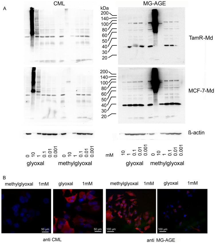
Figure 3. AGE accumulation under exogenous aldehyde stress.
A: AGE accumulation (CML (left) and MG-AGE (right)) in MCF-7-Md (lower panel) and TamR-Md cells (upper panel) after cultivation for three days with different concentrations of methylglyoxal and glyoxal as shown by Western blotting. Strongly enhanced AGE accumulation became visible when cell number was significantly reduced due to toxic effects, as represented by the decreasing β-actin signal. Glyoxal resulted in accumulation of CML whereas methylglyoxal treatment caused MG-AGE modification. Cells were treated as described for the determination of vitality/proliferation. Proteins were extracted per well and not corrected for protein amount. Therefore, the blots represent adherent cells only. B) AGE accumulation shown by immunofluorescence. Composite images of dual exposures are shown. MCF-7-Md cells were stained for CML and MG-AGE (red) by specific antibodies as indicated and nuclear staining was achieved with DAPI (blue) of cells treated for 24 h with 1 mM methylglyoxal or glyoxal.

