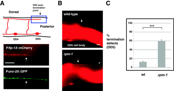
Figure 6.

rpm-1 regulates axon termination of the DD5 motor neuron. (A) Schematic highlights the axon termination site of the DD5 neuron (arrow) (inspired by Worm Atlas). Blue box highlights the region of the DD5 axon that was visualized using epifluorescent microscopy and two transgenes: Punc-25GFP (juIs76) and Pflp-13mCherry (bggIs6). mCherry highlights the DD5 termination point (arrow), while GFP fills both DD5 and DD6. (B) Arrow highlights the normal DD5 axon termination point. In rpm-1 mutants, the DD5 axon overextends (arrowhead). (C) Quantitation of DD5 axon termination defects for the indicated genotypes. For each genotype, the mean is shown from five or more counts (at least 20 worms/count). Analysis was performed on young adults grown at 23°C. Error bars represent the standard error of the mean. Significance was determined using an unpaired Student’s t test: ***P < 0.001. Scale bars, 10 μm. DD, dorsal D neuron; wt, wild-type.
