Figure 7.

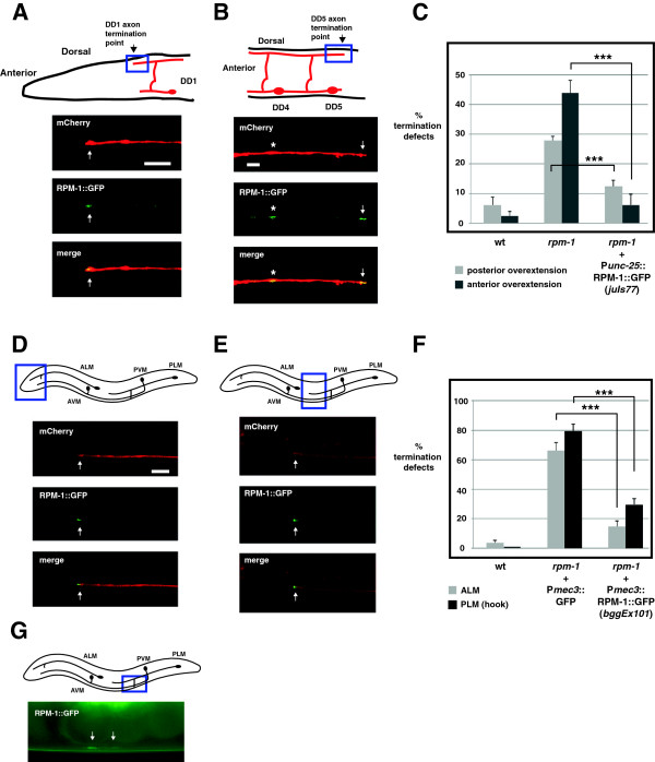
RPM-1 localizes to the axon tips and presynaptic terminals of GABAergic motor neurons and mechanosensory neurons. (A) Schematic shows the termination site of the DD1 neuron (arrow) (inspired by Worm Atlas). Blue box highlights where confocal microscopy was used to visualize Punc-25RPM-1::GFP (juIs77) and Pflp-13mCherry (bggEx99). RPM-1::GFP is concentrated at the tip of the DD1 axon (arrow). (B) Schematic shows the morphology and termination site of the DD5 neuron. Blue box highlights where confocal microscopy was used to visualize Punc-25RPM-1::GFP (juIs77) and Pflp-13mCherry (bggEx99). RPM-1::GFP is concentrated at presynaptic terminals (asterisk) and the tip of the DD5 axon (arrow). (C) Punc-25RPM-1::GFP (juIs77) rescues axon overextension defects caused by rpm-1 (lf) in the posterior and anterior tip of the dorsal cord. For each genotype, the mean is shown from five or more counts (at least 20 worms/count). (D,E) Schematic shows the mechanosensory neurons of C. elegans. Blue box highlights the region of the animal where confocal microscopy was used to visualize Pmec-7mCherry or Pmec-3RPM-1::GFP (bggEx101). RPM-1::GFP is concentrated at the tip (arrow) of (D) the ALM axon and (E) the PLM axon. (F) Pmec-3RPM-1::GFP (bggEx101) rescues axon termination defects in ALM (gray) and PLM neurons (hook, black bars). For each genotype, the mean is shown from three or more counts (at least 20 worms/count). For Pmec-3GFP (negative control), three independently derived transgenic lines were analyzed. (G) Blue box highlights the region of the PLM that was visualized by epifluorescent microscopy. Shown below is RPM-1::GFP (bggEx101) concentrated at the presynaptic terminals of a PLM neuron (arrows). All images and analysis were generated using young adult animals grown at 23°C. Error bars represent the standard error of the mean. Significance was determined using an unpaired Student’s t test: ***P < 0.001. ALM, anterior lateral microtubule; AVM, anterior ventral microtubule; PLM, posterior lateral microtubule; PVM, posterior ventral microtubule; wt, wild-type. Scale bars, 5 μm.
