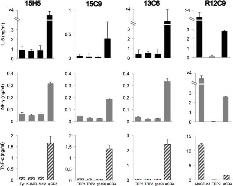
Figure 7.
Cytokine-secretion assay with selected clones stimulated with individual proteins. After induction of protein expression by Isopropyl β-D-thiogalactoside (IPTG) and opsonization with complement, the bacteria were added to autologous Epstein-Barr virus-transformed B cells (EBV-B cells) and incubated overnight. CD4 T cells (5,000/well) were added to EBV-B cells (30,000/well). The amount of tumor necrosis factor (TNF)-α, interferon (IFN)-γ, interleukin (IL)-5, and IL-17 in the supernatant of overnight co-cultures was estimated with a multicytokine bioplex kit. As the positive control, anti-MAGE-3 T cell clone R12C9 was used. The screening of the clones was performed in triplicates. Error bars represent SD.

