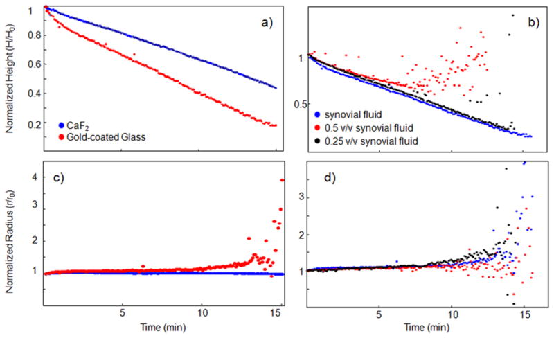
Figure 2.

The contact angle, drop height, and drop radius were calculated from side-profile images collected from evaporating human synovial fluid drops. We examined the effect of substrate surface (panels a and b) and biofluid concentration (panels c and d) on these geometric properties. Geometric properties were studied in normal synovial and degenerative joint condition (DJC) synovial fluid (shown in Supplementary Figure 2) deposited onto gold-coated glass and calcium fluoride. The radius of a synovial fluid drop remained consistent throughout the evaporation, indicating a fixed contact point, independent of substrate surface, shown in Supplemental Figure 2.. The drop height decreased uniformly in all experiments. From these data, the piling model of drop deposition could be applied toward synovial fluid.
