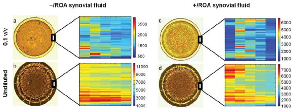
Figure 7.

Low magnification (4x) and Raman maps of the outer ring for −/ROA and +/ROA synovial fluid, at low concentration (0.1 v/v) and undiluted. Similar to plasma, concentration has an effect on synovial fluid drop deposition. Undiluted synovial fluid dried with a thick outer ring, with cracks radiating toward the outer edge. Diluted synovial fluid drops dried as ring-shaped deposits with a thin outer ring and no cracks. Raman maps were generated from the intensity sums of the 1003,1033,1206,1445,1655 cm−1 bands, attributed to represent protein content. Scale bars on the Raman maps reflect relative intensities, and should not be used to qualitatively compare protein levels between two different drops. Qualitative analysis of the Raman maps show that protein distribution within the outer ring is highly variable, even though the ring appeared smooth, and dependent on synovial fluid concentration. The greatest spatial heterogeneity was observed in drops of undiluted synovial fluid.
