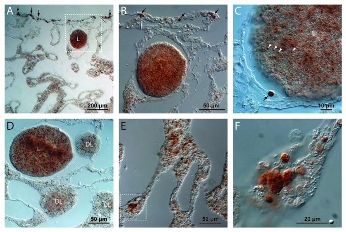
Figure 3. Immunocytochemistry reveals A. willeyana LC3A+ cells. (A) A low magnification image of a section illustrating the inner choanosome and the outer ectosome of A. willeyana. Near the outermost ectosome layer is a larva (L) containing LC3A-positive cells. Also clearly visible in this section are spherulite-forming ectosomal LC3A+ cells (vertical arrows). (B) A magnification of the boxed region in A detailing the LC3A+ cells in the larva (L), and the larger mature cells in the ectosome. (C) An adult LC3A+ cell (black arrow) in close proximity to a larva illustrates the clear difference in size between differentiated spherulite-forming cells and larval LC3A+ cells (white arrowheads). (D) Developing larvae (DL) and relatively mature larvae (L) contain LC3A+ cells suggesting that the process of autophagy is active throughout development in A. willeyana. However LC3A+ cells in larval stages do not have the same morphology as fully differentiated adult spherulite-forming cells, suggesting that these larval cells are not forming spherulites. (E) An image from the deeper choanosome layer where there can be many LC3A+ cells. (F) A magnified view of the boxed region in (E) illustrates the rounded morphology of these spherulite-forming cells.

An official website of the United States government
Here's how you know
Official websites use .gov
A
.gov website belongs to an official
government organization in the United States.
Secure .gov websites use HTTPS
A lock (
) or https:// means you've safely
connected to the .gov website. Share sensitive
information only on official, secure websites.
