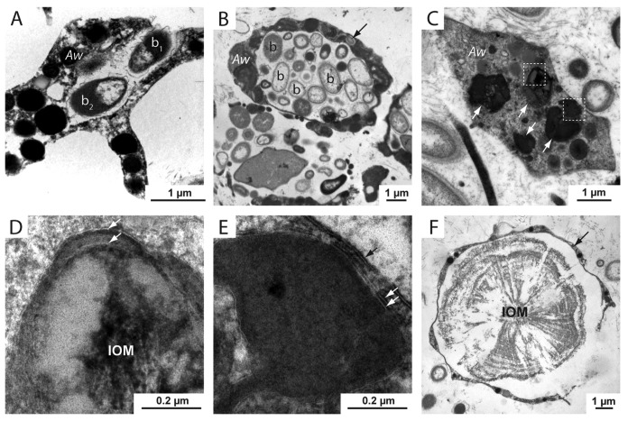
Figure 4. TEM sections of A. willeyana tissue reveal a rich population of inter- and intracellular bacteria, and subcellular structures bound by multiple membranes. (A) An A. willeyana cell (Aw) in the process of phagocytosing a bacterial cell (b1). (B) A representative TEM section illustrating the abundant intracellular bacterial community of A. willeyana. Indicated are bacterial cells (b) located within a large membrane-bound space within the sponge cell (Aw). A bacterial cell located within the cytoplasm is indicated with an arrow. (C) An A. willeyana cell (Aw) containing likely autophagosomes (white arrows). (D) Magnification of the upper left-most boxed region in (C). A double membrane is indicated by 2 white arrows, and the heterogeneous IOM that will form the material upon which calcification will take place is clearly visible. (E) Magnification of the lower boxed section in (C) illustrates another double membrane enclosing an autophagosome (white arrows). The cell membrane is indicated by a black arrow. (F) A TEM section of decalcified A. willeyana tissue. Shown is a spherulite-forming cell (arrow) surrounding the insoluble organic material (IOM) that is normally occluded within the calcified spherulite. Reproduced from reference 7.

An official website of the United States government
Here's how you know
Official websites use .gov
A
.gov website belongs to an official
government organization in the United States.
Secure .gov websites use HTTPS
A lock (
) or https:// means you've safely
connected to the .gov website. Share sensitive
information only on official, secure websites.
