
Fig. 3.
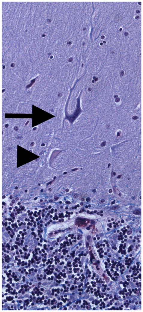
Luxol fast blue hematoxylin and eosin-stained cerebellar cortical section of an ET case showing a heterotopic Purkinje cell (long arrow). The Purkinje cell is oriented abnormally and a dendritic swelling is shown closer to the granular layer (i.e., beneath the cell, arrowhead). ×20 magnification