Figure 5.
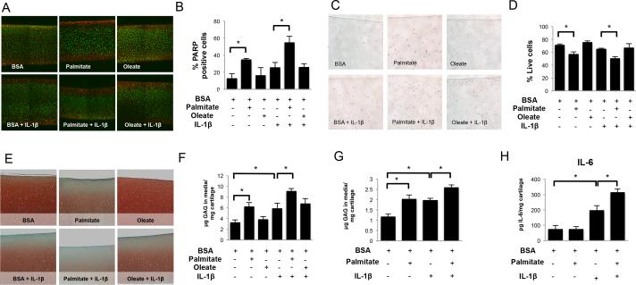
Free fatty acids effects in bovine and human cartilage explants. Full-thickness cartilage explants were treated with palmitate (0.5 mM), oleate (0.5 mM), or IL-1β (1 ng/ml) as indicated for 72 hours. (A) Chondrocyte viability visualized by live/dead assay and (B) percentage of live chondrocytes in bovine cartilage explants. Magnification: 40X. (C) Cleaved poly (ADP-ribose) polymerase (PARP) detection by immunohistochemistry and (D) percentage of PARP positive chondrocytes in bovine cartilage explants. Magnification 100X. (E) Safranin O staining of glycosaminoglycans (GAGs) and (F) GAG release into the supernatants in bovine cartilage explants. Magnification: 40X. (G) GAGs and (H) IL-6 concentration in the supernatants of cultured human cartilage explants. Images are representative of two different experiments performed in triplicate. Values are expressed as mean ± SEM. *, p<0.05.

