
Fig. 2. ALDH2 deficiency accelerates the ethanol-induced liver inflammatory response (eg. IL-6 production) via MAA-mediated activation of Kupffer cells. (A).
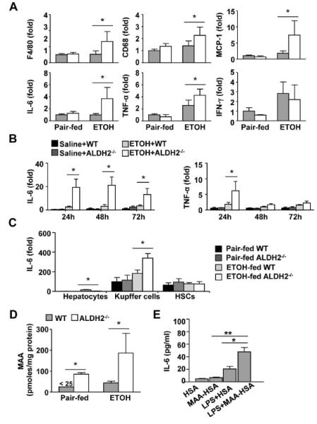
WT and ALDH2−/− mice were fed a control or ethanol diet for 10 days, followed by a single gavage of maltose or ethanol, respectively. Hepatic cytokine expression was measured using real-time PCR analyses. (n=4 in pair-fed WT or KO group, n=8 in ethanol-fed WT or KO group) (B) Expression of IL-6 and TNF-α mRNA in precision-cut liver slices (PCLS) ex vivo. (n=3 each group). (C) Hepatocytes, Kupffer cells, and HSCs were isolated from pair-fed or chronic-binge ethanol-fed mice and subjected to real-time PCR analyses. (n=3 each group). (D) Hepatic MAA levels from pair-fed or chronic-binge ethanol-fed mice were measured. (E) Kupffer cells from WT mice were isolated and treated with 25 μg/ml HSA or MAA-HSA in the presence or absence of LPS (1 ng/ml). After stimulation with MAA for 2 h, cell culture media were analyzed for IL-6 levels. (n=3 each group). The values represent means ± SD. *P < 0.05, **P < 0.01.