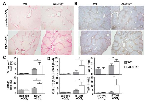
Fig. 6. ALDH2−/− mice show higher degrees of hepatic fibrosis after 8-week ethanol plus CCl4 treatment than do WT mice.

(A-D) WT and ALDH2−/− mice were treated with ethanol-fed plus CCl4 or pair-fed plus CCl4 for 8 weeks. (n=8 mice in each pair-fed or ethanol-fed WT or ALDH2−/− group). (A, B) Representative photographs of Sirius red staining (A) and immunohistochemical analysis with an anti-α-SMA antibody (B). (C) The Sirius red+ and α-SMA+ areas were quantified from panels A and B, respectively. (D) Real-time PCR analyses of hepatic fibrosis-associated genes. The values represent means ± SD. *P < 0.05.
