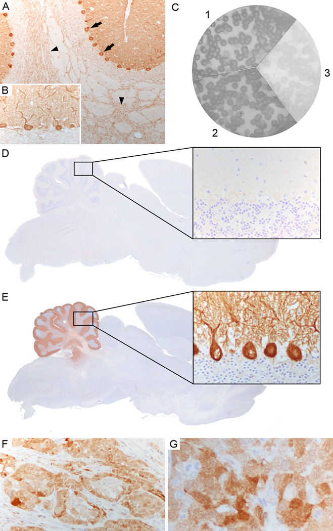
Figure 1. Characterization of CARP VIII antibody.
(A, B) Patient’s serum strongly labels Purkinje cell cytoplasm (arrows, A), axons (arrowheads, A), and dendrites (B) in rat cerebellum. (C) Filters with purified phage plaques expressing CARP VIII specifically reacted with a commercial CARP VIII antibody (1), and the patient’s serum (2) but not with serum from a healthy individual (3). (D, E) Competitive inhibition: Immunohistochemistry on rat brain, first incubated with the patient’s serum and subsequently labeled with a biotinylated CARP VIII antibody, is negative (D, rectangle in cerebellum enlarged in higher magnification) whereas immunohistochemistry on rat brain that was first incubated with a healthy control serum shows strong positive staining with the biotinylated CARP VIII antibody (E, rectangle in cerebellum enlarged in higher magnification). (F, G) Tumor biopsy of the ovarian adenocarcinoma shows expression of CARP VIII in the tumor cells. Magnification: A, F: x200; B, G, and enlarged rectangle in D, E: x400;

