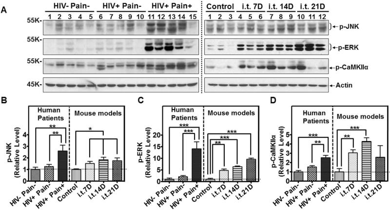
Figure 8.
Comparison of signaling pathways in the SDH of ‘pain-positive’ HIV-1 patients and i.t. gp120 mice. A. Immunoblots of phosphorylated JNK (p-JNK), phosphorylated ERK (p-ERK) and phosphorylated CaMKIIα (p-CaMKIIα) in the SDH of human patients (left panel) and i.t. gp120 mice at day 7 (7D), 14D or 21D (right panel). B-D. Quantitative summary of p-JNK (B), p-ERK (C), and p-CaMKIIα (D). (*, p<0.05; **, p<0.01; ***, p<0.001; One-way ANOVA)

