
Figure 4.
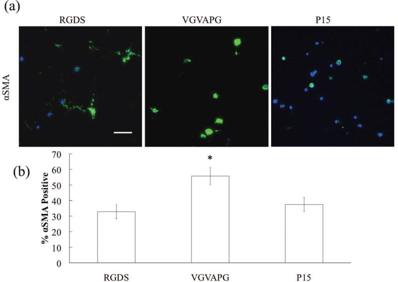
(a) Representative images of αSMA (green) immunostaining of VICs (nuclei in blue) after 14 days in culture. Scale bar = 100 μm. (b) Percentage of αSMA positive VICs as a function of adhesive small peptide after 14 days.
Official websites use .gov
A
.gov website belongs to an official
government organization in the United States.
Secure .gov websites use HTTPS
A lock (
) or https:// means you've safely
connected to the .gov website. Share sensitive
information only on official, secure websites.
(a) Representative images of αSMA (green) immunostaining of VICs (nuclei in blue) after 14 days in culture. Scale bar = 100 μm. (b) Percentage of αSMA positive VICs as a function of adhesive small peptide after 14 days.