Figure 6.
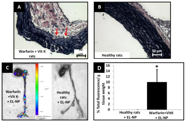
Targeting of EL-NPs in vascular calcification model (A) VVG histological stain for rat aorta after 3 weeks of warfarin + vitamin K (WK) treatment (arrow marks indicate fragmented elastin fibers, black) (B) healthy rat aorta. (C) IVIS imaging of whole heart and aorta at 24 hours after intravenous injection of EL-NPs. Strong fluorescent signal indicates the localization of ELNPs at specific sites of elastic lamina fragmentation. (D) Targeting of EL-NPs in healthy Vs. WK animals at 24 hours after intravenous injection showing significantly higher fluorescence in the aortic arch (n=6).

