
Figure 10.
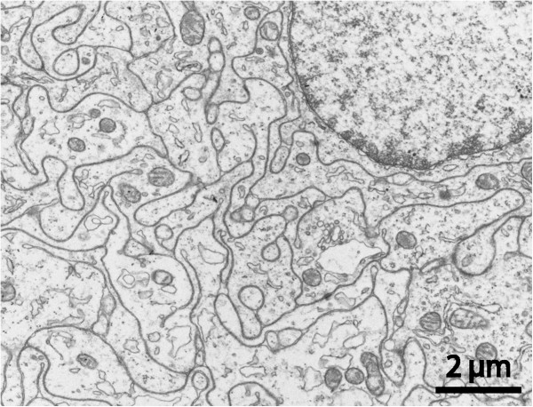
Aberrant-type cyst. TEM section from cyst wall with numerous tightly interlocking cell processes containing microtubules, filaments, scattered vesicles and some mitochondria, i.e. exhibiting neuropil morphology. Occasional myelinated fibers were encountered (not shown).