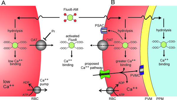
Figure 7.

Schematic showing proposed Ca++and Fluo-8 transport mechanisms. (A) Uninfected human erythrocytes maintain a low intracellular Ca++ through a low passive Ca++ permeability and an ATP-dependent extrusion pump (Ca++ pump). Fluo-8 AM enters cells by diffusion and is hydrolyzed to yield activated Fluo-8, which fluoresces upon Ca++ binding. Fluo-8 may be exported via organic anion transporters (OAT). (B) Infected cells have a higher intracellular Ca++ through activation of a distinct Ca++ uptake pathway. Ca++ that enters the cell may be exported via the Ca++ pump, may remain in the host cytosol, or may enter parasite compartments by crossing the parasitophorous vacuolar membrane (PVM) through nonselective PVM channels (PVMC) [39,40] and the parasite plasma membrane (PPM) via unknown mechanisms. Fluo-8 AM that enters infected cells may be hydrolyzed to the active form in various compartments; the activated dye may be exported via the host cell OAT and possibly via PSAC. Transport of the dye across parasite membranes may also occur, but is not illustrated.
