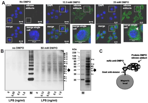
Figure 4. Localization and identification of protein-DMPO nitrone adducts in RAW 264.7 macrophages treated with LPS and DMPO.
A) Single-plane confocal images of nitrone adducts formed in cells treated with 1 ng/ml LPS and 12.5 or 50 mM DMPO for 24 h. Green indicates nitrone adducts and blue indicates nuclei. Insert is a high-power magnification of the image of a single and representative cell. The white arrowhead indicates the perinuclear localization of most nitrone adducts. B) Western blot analysis of protein-DMPO nitrone adducts in homogenates of cells treated with LPS and/or DMPO for 24 h. Right panel, coomassie blue staining of the homogenate of cells treated with 1 ng/ml LPS and 50 mM DMPO for 24 h, separated in a reducing gel and showing 7 representative bands that correspond to anti-DMPO-positive bands in the Western blot. C) Schematic representation of an anti-DMPO molecular “catcher”-protein-DMPO nitrone adduct complex used to pull-down proteins labeled with DMPO. M indicates Magic Mark Western XP molecular weight marker (Invitrogen). Modified from [29].

