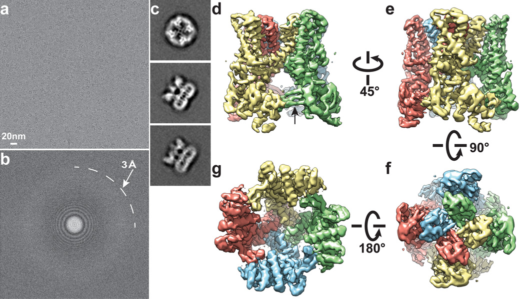
Figure 1. 3D reconstruction of TRPV1 determined by single-particle cryo-EM.
a, Representative electron micrograph of TRPV1 protein embedded in a thin layer of vitreous ice recorded at a defocus of 1.7µm. b, Fourier transform of micrograph shown in a, with Thon rings extending to nearly 3Å. c, Enlarged views of three representative 2D class averages show fine features of tetrameric channel complex. d–g, 3D density map of TRPV1 channel filtered to a resolution of 3.4Å (scaled to atomic structure) with each subunit color-coded. Four different views of the channel are shown, from side (d, e), top (f), and bottom (g). The arrow in panel d indicates β-sheet structure in the cytosolic domain of TRPV1.

