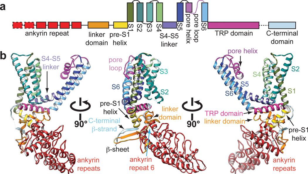
Figure 3. Structural details of a single TRPV1 subunit.
a, Linear diagram depicting major structural domains in a TRPV1 subunit, color coded to match ribbon diagrams below. Dashed boxes denote regions for which density was not observed (first two ankyrin repeats) or where specific residues could not be definitively assigned (C-terminal β-strand). b, Ribbon diagrams showing three different views of a TRPV1 monomer denoting specific domains.

