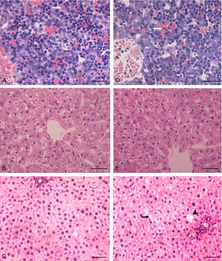
Fig. 2.
Evidence of hepatic steatosis in adult IUGR offsprings. HE staining was performed for the sections from rat livers at different developmental stages. (A)–(C) show representative sections from CON rats (40× original magnification) (A) E20, (B) PW3, (C) PW40. (D)–(F) show sections from IUGR rats (D) E20, (E) PW3, (F) PW40. White arrow indicates immature erythrocytes; black arrow indicates microvesicular fat droplets; black arrow head indicates macrovesicular fat droplet. Scale bars = 50 µm.

