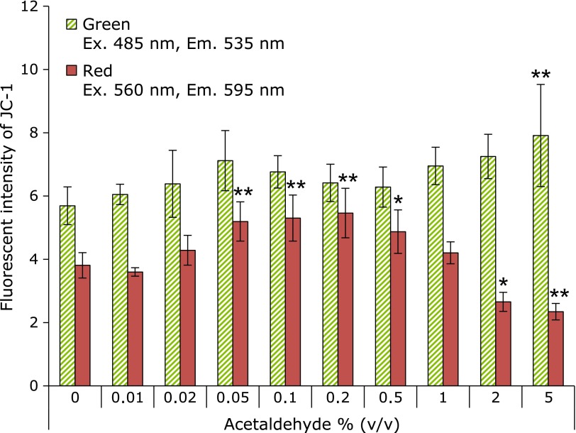
Fig. 5.
Mitochondrial injury by exposing acetaldehyde. Mitochondrial injury was measured by JC-1 after exposed-ethanol for 1 h. Green fluorescence (Ex. 485 nm, Em. 535 nm) and red fluorescence (Ex. 560 nm, Em. 595 nm) show mitochondrial injury and healthy mitochondria, respectively. The fluorescence intensity was measured by plate reader. 0.5–5% acetaldehyde-exposed cells were completely death. n = 6, Error bar; SD. *p<0.05, **p<0.01.

