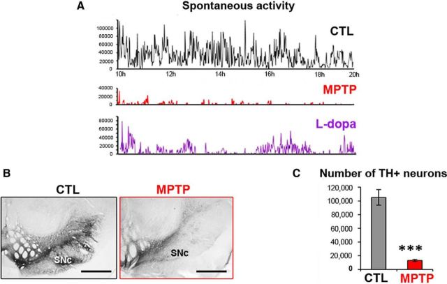
Figure 2.
Measurement of motor disability and assessment of DA lesion within the mesencephalon of MPTP-treated macaques. A, Examples of 10 h daytime recording of spontaneous activity of MPTP-treated Macaque 3 during the control (CTL) state, after MPTP intoxication, then after l-dopa medication. l-Dopa treatment (bottom graph) was given at 10 h and at 14 h. B, Example of TH immunostaining in the mesencephalon of a control macaque and an MPTP-treated macaque. Scale bar, 2 mm. C, Graphic representation of the number of TH+ neurons in the substantia nigra pars compacta (SNc) in the 4 MPTP-treated macaques and 5 controls. ***p < 0.001 (Mann–Whitney rank-sum test).

