
Figure 4.
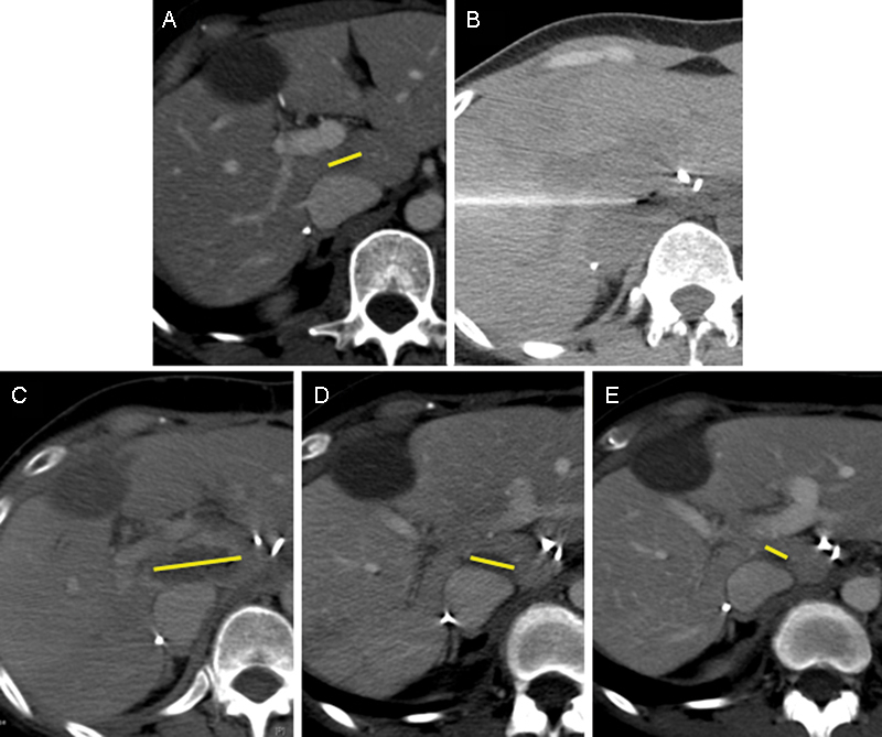
CT imaging of a 48-year-old woman with metastatic colon cancer to the liver. (A) Pretreatment tumor imaging (yellow bar shows major axis of tumor). (B) Treatment CT image after placement of two monopolar electrodes in the liver. (C) Immediate postprocedure imaging demonstrating hypodense ablation zone (yellow bar shows major axis of ablation zone). (D) One-month postablation imaging showing an involution of the ablation cavity (yellow bar shows major axis of ablation zone). (E) Four-month postablation imaging showing continued involution of the ablation cavity (yellow bar shows major axis of ablation zone). CT, computed tomography.