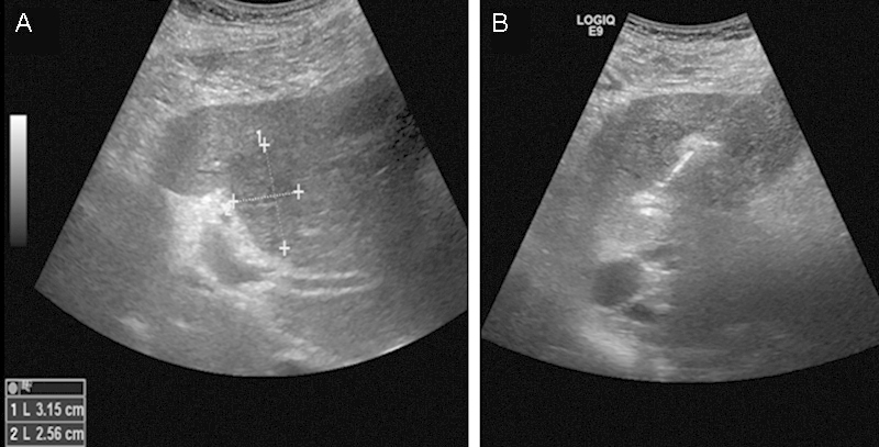
Figure 5.

Ultrasound images during IRE of a 76-year-old man with metastatic colon cancer to the liver. (A) Targeted tumor dimensions. (B) Image obtained half way through the treatment. The area treated by IRE becomes hyperechoic compared with surrounding liver tissue. IRE, irreversible electroporation.
