Figure 5.
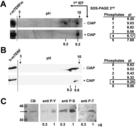
mTERF is phosphorylated at multiple sites. (A) Two-dimensional gel electrophoresis (2D) of in vitro synthesized and [35S]methionine-labeled rat mTERFm before (–) and after (+) treatment with CIAP. (B) Another similar experiment carried out with human mtS100 lysate (human mTERF) and revealed with a polyclonal antibody anti-mTERF. Both methods showed a phosphorylated spot at pI ∼8.2 for rat mTERFm and ∼9.2 for human mTERF, which correspond to four phosphorylated residues (right panel). The left sides show rat mTERFm and human mTERF when they were running in a well in parallel in SDS–PAGE. (C) The bacteria expressed and purified rat his-mTERF was revealed using antibodies against phosphotyrosine (anti P-Y), phosphoserine (anti P-S) and phosphothreonine (anti P-T). CB was stained with Coomassie blue.

