
Figure 10.
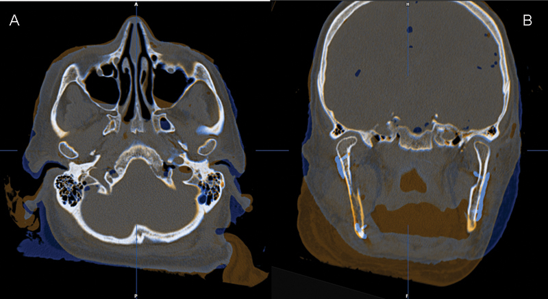
The fusion of preoperative (amber) and postoperative (blue) CT datasets demonstrated that the positions of the two residual rami and condyles after the fixation of the CAD/CAM-fabricated reconstruction plate were almost identical to those before resection. (A) Axial view. (B) Coronal view.