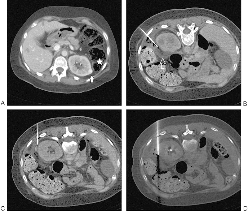
Figure 2.

(A) A 38-year-old woman with tuberous sclerosis and prior right nephrectomy for renal cell carcinoma found to have a 1.1-cm enhancing left renal mass (arrow). Note the immediately adjacent colon (star). (B) Insertion of 21-gauge needle allows hydrodissection to be performed with injection of sterile water (arrow) to displace the adjacent colon away from the targeted lesion. (C) RFA electrode positioned under CT guidance, just prior to final position across the lesion. (D) RFA electrode in final position across the lesion with colon successfully displaced. CT, computed tomography; RFA, radiofrequency ablation.
