
Fig. 1.
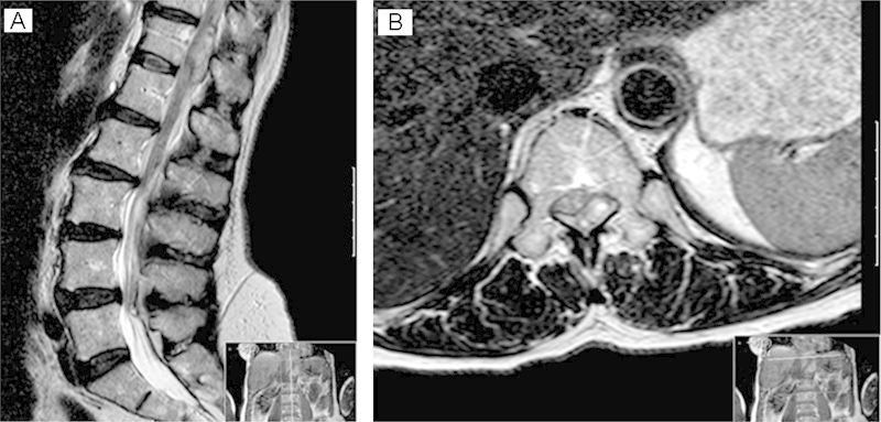
(A, B) Sagittal and axial magnetic resonance image of the thoracolumbar spine showing an extra axial space-occupying lesion compressing the cord at the level of T12. This appears to be occupying epidural space and is posterior and to the left of the theca. There is high signal within the adjacent cord consistent with a compressive myelopathy.