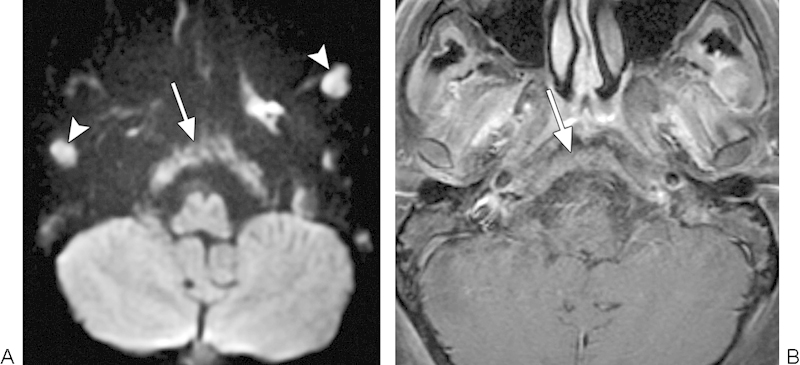
Fig. 15.

Metastases. (A) Diffusion-weighted imaging shows diffuse involvement of the clivus by metastatic disease (arrow). There are additional metastatic lesions within the bilateral masticator spaces (arrowheads). (B) The clivus lesion (arrow) is less conspicuous on the corresponding postcontrast T1-weighted image.
