
Fig. 3.
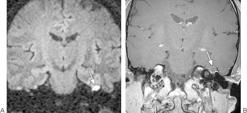
Recurrent cholesteatoma. (A) Coronal turbo spin-echo diffusion-weighted image shows a hyperintense focus within the right mastoid bowl (arrow). (B) Coronal postcontrast T1-weighted magnetic resonance imaging shows that the lesion does not enhance (arrow).