Fig. 9.

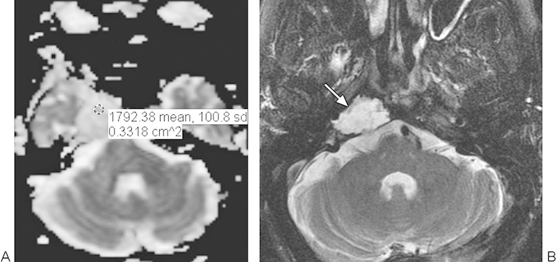
Low-grade chondrosarcoma. (A) Apparent diffusion coefficient map with a sample region of interest shows high diffusivity throughout the tumor. (B) T2-weighted magnetic resonance imaging shows homogeneous high signal in the mass (arrow).

Low-grade chondrosarcoma. (A) Apparent diffusion coefficient map with a sample region of interest shows high diffusivity throughout the tumor. (B) T2-weighted magnetic resonance imaging shows homogeneous high signal in the mass (arrow).