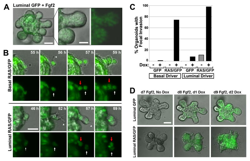
Figure 3. H-RASG12V, but not Fgf2, triggers focal invasion into Matrigel.
(A) Fgf2-induced multilayered epithelial elongation. Still image from a live cell imaging experiment depict a representative luminalGFP organoid 7 d after initiating Fgf2-induced branching morphogenesis. (B) Ras-mediated focal invasion. Time-lapse images depict leader cells initiating invasion. Arrowheads mark protrusions emanating from leader cells indicated by white arrows. Red Arrows indicate follower “stalk” cells. Bar, 25μm. (C) Quantification of Ras-mediated focal invasion. Organoids of the indicated genotypes were cultured in the presence and absence of Dox for 6 d, then scored for the presence of focal invasion as described in Materials and Methods. Mean values reflect analysis of 20 to 77 organoids for each genotype and treatment condition. (D) Contrasting modes of collective MEC invasion triggered by Fgf2 versus H-RASG12V. Organoids of the indicated genotypes underwent Fgf2-induced branching prior to Dox treatment. Panels depict morphology of a representative organoid before and after Dox-induced transgene expression.

