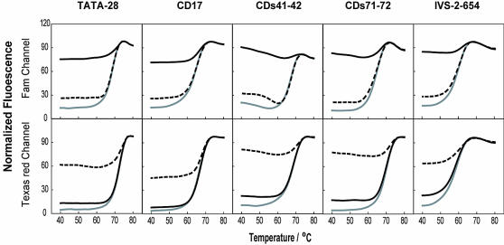
Figure 3.
Thermal denaturation curves of the five pairs of displacing probes used for HBB genotyping. The fluorescence intensity of solutions containing wild-type probes (upper panel) and mutant probes (lower panel) was plotted as a function of temperature in the absence of targets (gray solid lines), in the presence of the wild-type target (black solid lines) and in the presence of the mutant targets (dashed lines). The wild-type targets are oligonucleotides perfectly complementary to the positive strand of the wild-type probes, and the mutant targets are oligonucleotides perfectly complementary to the positive strand of the mutant-type probes.

