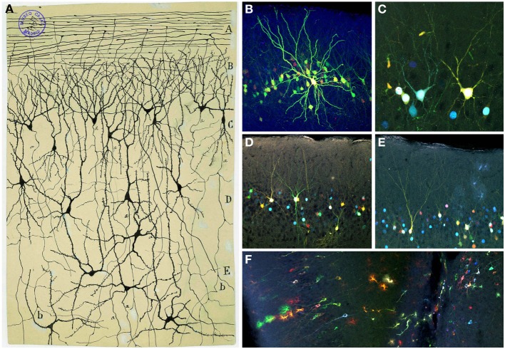
Figure 11.
Olfactory cortex. (A) Original Cajal drawing showing the olfactory cortex layers (Ramón y Cajal, 1901). Olfactory fibers layer (A); plexiform layer (B); layer of polymorphic superficial cells (C); layer of the pyramids (D); deep polymorphous cells (D). (b) Bifurcation of axons. Cajal Legacy (Instituto Cajal, CSIC, Madrid, Spain). (B–F) Different cell morphologies in the adult mouse olfactory cortex labeled after E12 in utero electroporation of different plasmids with the UbC-Star Track method (Figueres-Oñate and López-Mascaraque, 2013). Note the presence of cells with either arachnoid morphologies, similar to those in (B), and crescent-shaped cells similar to (C). (D,E). View of different morphological neuronal types. (F) Several neurons along with different glial clones.

