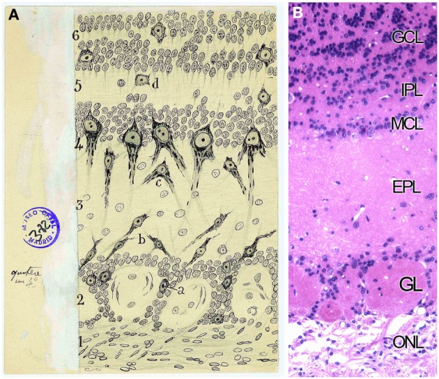
Figure 5.

Layers distribution of the Olfactory bulb. (A) Original Cajal drawing of the frontal section of the rabbit olfactory bulb (Ramón y Cajal, 1901). (1) Nerve layer (ONL). (2) Glomerular layer (GL). (3) External plexiform layer (EPL). (4) Mitral cells layer (MCL). (5) Inner plexiform layer (IPL). (6) Grains and white matter layer (GcL). (a) Peripheral tufted cells; (b) middle; (c) internal (d) short axon cells. Cajal Legacy (Instituto Cajal, CSIC, Madrid, Spain). (B) Olfactory bulb mouse coronal section of hematoxylin and eosin nuclei staining showing the different layers described by Cajal in (A).
