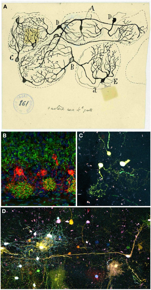
Figure 6.

Periglomerular cells. (A) Original drawing by Cajal's disciple Blanes, showing periglomerular cells of a cat glomerular layer (Blanes, 1898). Cell branching into three glomeruli (A). Biglomerular cells (B–D). Monoglomerular cells (C–E). (a) Axon. Cajal Legacy (Instituto Cajal, CSIC, Madrid, Spain). (B) Periglomerular cells immunolabeled for tyrosine hydroxylase (TH, red) protein and Dab1 protein (green) on mouse olfactory bulb sections at P3. Panel was taken by Eduardo Martin-López. (C,D) Adult periglomerular cells contributing to one (C) or more glomeruli (D) labeled after E13 in utero electroporation of different plasmids with the UbC-Star Track method (Figueres-Oñate and López-Mascaraque, 2013).
