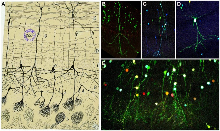
Figure 8.
Granular cells. (A) Original Cajal drawing of an olfactory bulb section from a few days cat brain (Ramón y Cajal, 1901). Glomerular layer (A), outer plexiform layer (B), mitral cell layer (C), inner plexiform layer (D), grains layer and white matter (E). (a) Terminal arborization of an olfactory fiber; (b) glomerulus with several endings; (c) mitral plume; (d) tufted cells. Cajal Legacy (Instituto Cajal, CSIC, Madrid, Spain). (B–E) Granule cells in the olfactory bulb of young adult mice (P20) labeled after E12-14 in utero electroporation of different plasmids with the UbC-Star Track method. (Figueres-Oñate and López-Mascaraque, 2013). (B,C) Granule cells with similar to (i,j) in Cajal's drawing. (D) Tufted cell. (E) Granular cells branching their processes in the glomeruli.

