Figure 1.
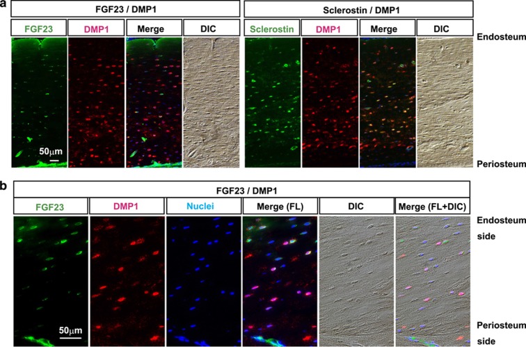
Distinct localization of FGF23 and DMP1 in cortical osteocytes. (a) Double immunofluorescence stainings for FGF23/DMP1 and sclerostin/DMP1 were performed with femur sections obtained from 16-week-old rats. FGF23 and DMP1 were detected by secondary antibodies conjugated with Alexa Fluor 488 (in green) and 568 (in red), respectively. The sections were counterstained with 4′-6-diamidino-2-phenylindole to detect nuclei (in blue). Differential interference contrast (DIC) images of each section were obtained for morphological observation. Merged images are shown in the right panels. Scale bar: 50 μm. Endosteal side is up. (b) Magnified images of double immunofluorescence stainings for FGF23/DMP1 of another specimen from a 16-week-old rat. Scale bar: 50 μm. Endosteal side is up. FL; fluorescence.

