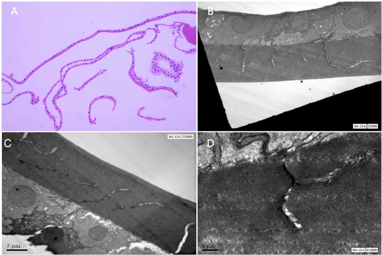
Figure 2.
Histopathology of the excised anterior capsule. (A) Markedly thinned lens capsule with atrophic epithelium in central portion (H&E, ×100). (B) EM of anterior capsule showed multiple linear and irregular zones of capsular dehiscence in inner two-third of capsule. (C) A normal lens epithelium with thinned anterior lens capsule. (D) A few of this dehiscence had a fibrillar and irregular electron dense material and vacuoles.

