Figure 1.

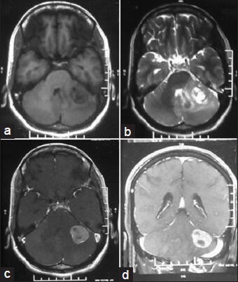
Magnetic resonance imaging scan showing a heterogeneous intra axial mass in left cerebellar hemisphere with T1 hypointensity, (a) T2 hyperintensity, (b) with contrast enhancement, (c) and coronal section showing same tumoral aspect (d)

Magnetic resonance imaging scan showing a heterogeneous intra axial mass in left cerebellar hemisphere with T1 hypointensity, (a) T2 hyperintensity, (b) with contrast enhancement, (c) and coronal section showing same tumoral aspect (d)