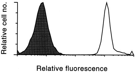
Figure 4.
Dansyl labeling of CHO cells. Fluorescent BSA-dansyl conjugate was coupled to the CHO cell surface, and the cells were analyzed by flow cytometry (excitation, 340 nm; emission, 565 nm). The relative fluorescence of cells coupled with BSA alone (shaded area) and dansylated BSA (unshaded area) is shown. CHO, Chinese hamster ovary.

