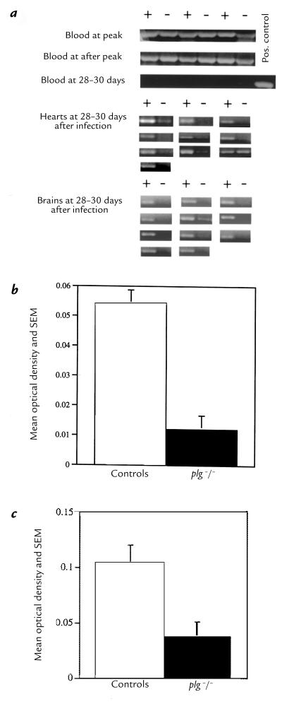
Figure 3.
PCR amplifications of spirochetal DNA (flaB) from blood and brains. (a) Representative amplifications of spirochetal DNA of blood from control (+) and plg–/– mice (–) obtained at peak, after peak, and 28–30 days after inoculation. Control and plg–/– mice (10 pair) were used to detect spirochetal DNA in hearts at 28–30 days. Control and plg–/– mice (11 pair) were used to detect spirochetal DNA in brains at 28–30 days. (b) Mean OD of the amplimers from the PCRs of the hearts of 10 pair of controls and plg–/– mice. (c) Mean OD of the amplimers from the PCRs of the brains of 11 pair of controls and plg–/– mice. For b and c, the mean OD of the uninfected PCR tissue controls was subtracted from the OD of each infected mouse.

