Figure 1.
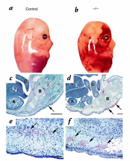
SLP-76–deficient mice manifest diffuse, subcutaneous hemorrhage and edema. SLP-76+/– mice were mated and day of gestation was calculated based on the presence of a vaginal plug. At approximately day 14 (a and b) or day 18 (c–f) of gestation, the mother was sacrificed and fetuses were isolated. Genotypes were determined by PCR analysis using genomic DNA isolated from a small tissue sample as template. (a and b) Gross morphological appearance of littermate control (a) or SLP-76–/– (b) E14 fetuses. (c–f) Histological appearance of SLP-76+/– (c and e) and SLP-76–/– (d and f) fetal sections at day 18 of gestation. (c and d) Caudal view of embryos. Note close association of epithelium with underlying connective tissue (arrow) in the SLP-76+/– fetus and edema and subcutaneous bleeding in the SLP-76–/– fetus. B, bone; G, gut; K, kidney; L, liver. Bar represents 1000 μM. (e and f) High-power view of subcutaneous region. Note intact endothelium and intraluminal nucleated red blood cells (arrows) in SLP-76+/– embryo compared with attenuated endothelium, extravasated blood (arrow), and subcutaneous edema in the SLP-76–/– embryo. Bar represents 200 μM.

