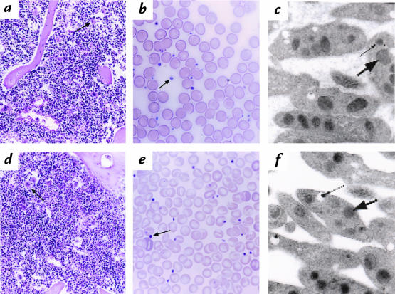
Figure 2.
Platelets and megakaryocytes develop normally and exhibit normal morphology in the absence of SLP-76. The humerus and peripheral blood were isolated from SLP-76+/+ (a–c) or SLP-76–/– (d–f) mice. Bone marrow sections (a and d), whole blood smears (b and e), or glutaraldehyde-fixed platelet sections were generated as described in Methods. (a and d) Hematoxylin and eosin–stained humeral bone marrow sections (×100). Megakaryocytes are indicated with arrows. Note that the density of megakaryocytes in sections obtained from SLP-76+/+ and SLP-76–/– mice was similar (1.1–1.15 megakaryocytes per oil immersion field). (b and e) Wright-Giemsa staining of whole blood smears (×250). Platelets are indicated with arrows. (c and f) Transmission electron microscopic analysis of glutaraldehyde-fixed platelets. Large arrows indicate α granules and small arrows denote dense granules.

