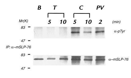
Figure 3.
SLP-76 is tyrosine-phosphorylated in response to collagen. Platelets were isolated from normal mice and left untreated (basal, B) or incubated with thrombin (T), collagen (C), or pervanadate (PV) for the times indicated. Platelets were then lysed and subjected to immunoprecipitation with a murine SLP-76–specific antibody. Immunoprecipitates were washed, resolved by SDS-PAGE, transferred to nitrocellulose, and then immunoblotted with the phosphotyrosine-specific antibody 4G10 (α-pTyr). The immunoblot shown in the top panel was stripped and reblotted with an SLP-76–specific antibody (SLP-76) to demonstrate equal amounts of immunoprecipitated SLP-76 in each lane. Identical results were obtained in a separate experiment.

