Figure 1.
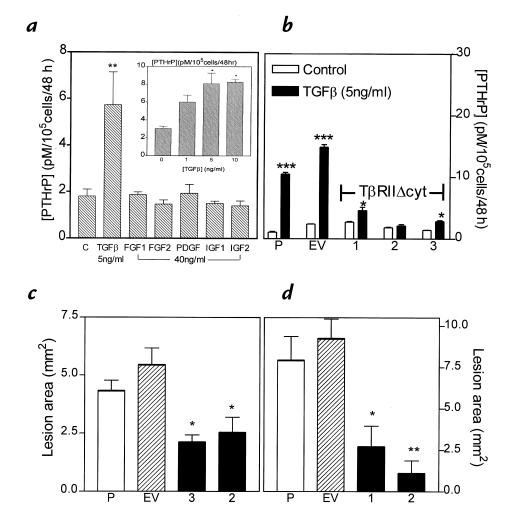
(a) Effects of bone growth factors on PTHrP secretion from MDA-MB-231 cells in vitro. MDA-MB-231 cells were plated onto 48-well plates and grown to near confluence. Cells were washed and treated with serum-free media containing the respective growth factors for 48 h. PTHrP concentrations in conditioned media were corrected for cell number. Only the results for the highest concentration of each growth factor are shown. Inset: Dose response for PTHrP secretion by MDA-MB-231 cells treated with TGF-β. Values represent the mean ± SEM (n = 3 per group).(b) Effect of TGF-β on PTHrP secretion by MDA-MB-231, MDA/pcDNA3, and MDA/TβRIIΔcyt clones. Respective cells were plated onto 48-well plates and treated as described in a. Values represent the mean ± SEM (n = 3 per group). P = parental MDA-MB-231; EV = empty vector pcDNA3 clone; 1, 2, and 3 are respective MDA/TβRIIΔcyt clones. (c and d) Osteolytic lesion area from radiographs of two separate experiments comparing clones 3 and 2 (c) or clones 1 and 2 (d) with controls of MDA-MB-231 (P) or pcDNA3 vector (EV). Values represent mean ± SEM (n = 4 per group). *P < 0.05, **P < 0.01, ***P < 0.001 vs. controls. PTHrP, parathyroid hormone–related protein; TGF-β, transforming growth factor-β.

