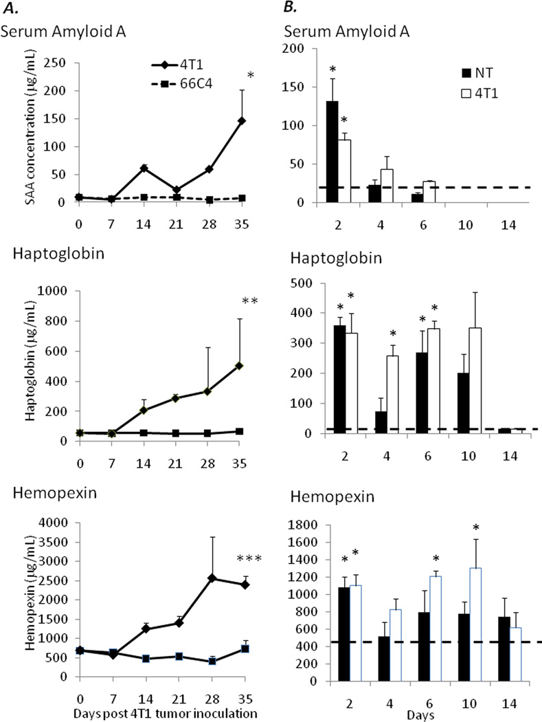
Figure 3.
(A) Plasma was harvested from 4T1- and 66C4-bearing mice at 0, 7, 14, 21, 28, and 35 days post tumor inoculation and assayed for serum amyloid A and haptoglobin via ELISA. Values represent mean concentration in ng/mL and are ± SD (n=4–5/group) (* Mann- Whitney Rank Sum Test p=0.01) (** Mann Whitney Rank Sum Test p=0.002) (*** Student’s t-test p <0.001). (B) Naïve Balb/c animals were injected intravenously with 1 × 10⁷ magnetically enriched Gr-1+ cells from healthy, nontumor and 4T1-bearing animals at days 1, 3, and 5. Serum was collected at days 2, 4, 6, 10, and 14 and assayed for concentrations of serum amyloid A, haptoglobin, and hemopexin. Hashed lines represent values obtained from sham injections (* p<0.05).

