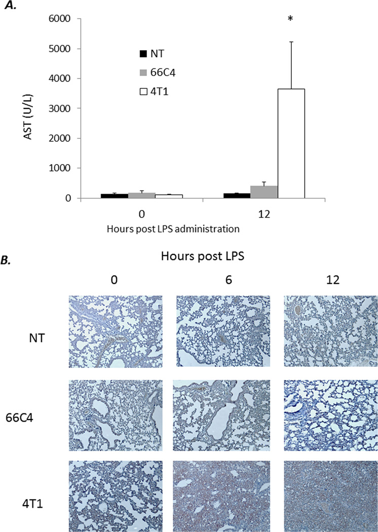
Figure 6. Overwhelming MDSC infiltration into lung and liver parenchyma following sublethal endotoxemia.
(A) Lungs were harvested from 4T1-, 66C4-, and nontumor- (NT) bearing mice at 0, 6, and 12 hours following sublethal endotoxin adminsitration, fixed in formalin, and stained with hematoxylin and eosin. Images taken at 10×. (B) Lung tissue was harvested from 4T1, 66C4, and NT bearing animals at 0 and 12 hours post LPS challenge and placed in formalin. Tissues were then sectioned and stained with hematoxylin and eosin. Arrows denote zone of necrosis. Images wre taken at 10×. (C) AST serum concentrations from 4T1-, 66C4-, and nontumor- (NT) bearing mice at 0 and 12 hours (*n=5 per group; Tukey’s One way ANOVA p<0.05).

